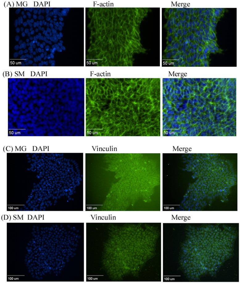
Figure 4. Organization of cytoskeletal structure proteins in hiPSCs grown on different surfaces.
(A, C, E): MG surface; (B, D, F): SM surface. hiPSCs were fixed after growing on the corresponding surfaces for two days and stained with antibodies against F-actin, vinculin, and zyxin, respectively. Arrows indicate denser and more pronounced actin filament expression. Scale bars: (A–B, E–F) 50 µm, (C–D) 100 µm. MG, Matrigel; SM, Synthemax Surface.

