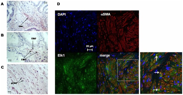
Figure 1. Elk1 expression in human prostate tissue.
(A), (B) Peroxidase staining of prostate tissues for Elk1. (A) Cytosolic Elk1 immunoreactivity in smooth muscle cells (smc). (B) Elk1-positive (Elk1+) and –negative (Elk1−) nuclei. (C) Peroxidase staining of prostate tissue for phospho-Elk1, with phospho-Elk-positive (pElk1+) nuclei. (D) Double fluorescence staining of human prostate tissue for Elk1 and αSMA. Yellow color in merged pictures represents Elk1 expression in smooth muscle cells. Shown are representative pictures from stainings from tissues of n = 6 patients for each staining.

