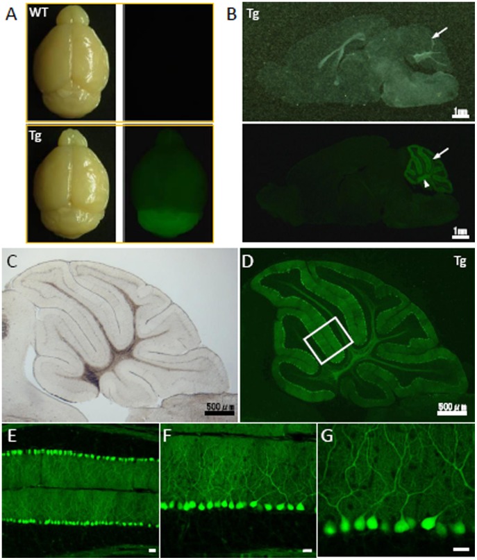
Figure 5. Cerebellum-restricted expression of GFP in the C57BL/6 MSCV-GFP mouse line.
(A) Brightfield (left) and fluorescent (right) images of the brains of transgenic mice (lower) and their wild-type littermates (upper). A high level of GFP expression was observed in the C57BL/6 MSCV-GFP mouse cerebellum. (B) A sagittal section of a C57BL/6 MSCV-GFP mouse brain is shown, again showing the cerebellum-selective expression of GFP. Scale bar, 1 mm. (C–G) Purkinje cell-specific expression of GFP in the C57BL/6 MSCV-GFP mouse cerebellum shown by brightfield (C) and fluorescent images (D). The boxed area in (D) is enlarged (E–G) to show the Purkinje cell-specific expression of GFP. Scale bar, 20 µm.

