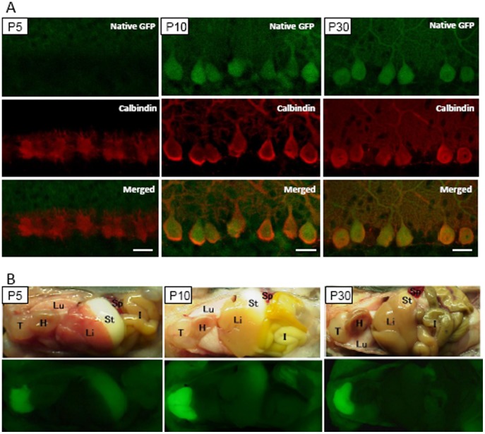
Figure 7. Expression of GFP during the development of C57BL/6 MSCV-GFP mice.
(A) No GFP expression was observed in the Purkinje cells at P5. Cerebellar sections obtained from MSCV-GFP mice at P5 (left), P10 (middle) and P30 (right) were immunolabeled for calbindin. Images of native GFP fluorescence (upper), calbindin immunoreactivity (middle) and merged images (lower) are presented. (B) Brightfield (upper) and fluorescent (lower) images of MSCV-GFP mice at P5, P10 and P30. Weak, but clearly detectable, levels of GFP were observed in the thymus from as early as P5 and increased thereafter. GFP fluorescence in the skeletal muscles became overt at P10. T; thymus, H; heart, Lu; lung, Li; liver, I; intestine, St; stomach, Sp; spleen.

