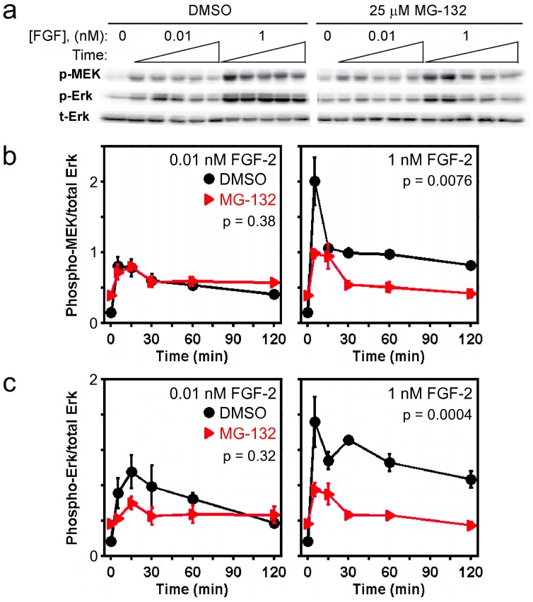
Figure 2. MG132 treatment reduces FGF-stimulated phosphorylation of MEK and ERK.
a) Immunoblot results, representative of 3 independent experiments, showing the kinetics of pMEK and pERK in cells pretreated with either DMSO or 25 µM MG132 for 6 h and then stimulated with the indicated concentration of FGF-2. Stimulation times are 5, 15, 30, 60, and 120 minutes. Total ERK1/2 (tERK) serves as a loading control. For each antigen, the DMSO and MG132 bands are cropped from the same gel. b&c) Quantification of the MEK (b) and ERK (c) phosphorylation kinetics represented in a. Each readout is normalized by total ERK and expressed as mean ± s.e.m. (n = 3). The indicated p value for each time course is from two-way ANOVA analysis comparing MG132-treated and control measurements.

