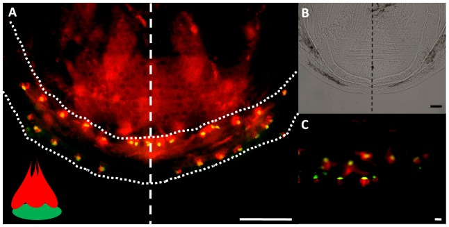
Figure 6. Immunohistochemical visualization of taste buds on the lower jaw at 21 dpf.
(A) Flat mounted lower jaw (outlined in dotted line) showing the inner and outer rows of taste buds. A dashed line separates the control side from the surgery side. Each taste bud is visualized as one green basal cell grouped with one or more red receptor cells, as depicted in the schematic inset in (A). (B) Is a bright field image of the jaw in (A) at a lower magnification; (C) shows a higher magnification of the taste buds. All scale bars are 50 µm.

