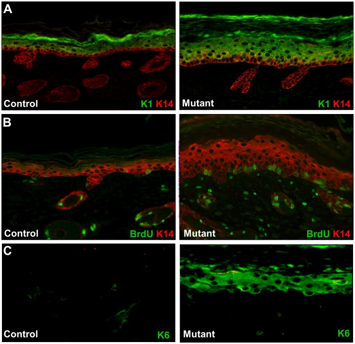
Figure 3. Hyperproliferation and altered expression of keratin markers.
A. Immunostaining with anti-K14 (red) and anti-K1 (green) antibodies. K14 expression in the control epidermis is predominantly in the stratum basale of the epidermis (left panel). In the mutant epidermis, K14 was detected in both basal and suprabasal layers (red and yellow color in right panel and panel B). Suprabasal differentiation marker K1 was detected in suprabasal cells of both mutant and control skin. The suprabasal layer in the mutant was thicker than the control. B. BrdU incorporation (green) and keratin K14 expression (red) were visualized by immunostaining. The mutant epidermis showed more than twice as many BrdU-staining cells in the basal epithelium. C. Immunostaining for K6 (green). K6 is not detected in the control epidermis (left panel), but K6 is strongly expressed in the suprabasal cells in the mutant. Original magnifications: X200.

