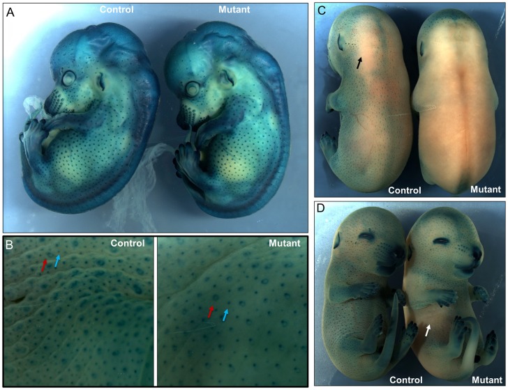
Figure 6. Alteration of skin structure at the earlier stages of pigskin mutants.
Hair follicle induction was assayed using a BMP4-lacZ reporter line from E14.5 to E16.5. Embryos were incubated in solution with 5-bromo-4-chloro-3-indolyl-b- D-galactoside (X-gal). X-gal cleavage by beta-galactosidase results in dark blue staining. A. At E14.5, representative control and pigskin mutant embryos display blue-stained primary hair follicles (PHFs) and vibrissal follicles. There were similar numbers of PHFs in the lateral side of control and mutant mice. B. Peeled skin from representative control and pigskin E16.5 embryos was stained with X-gal. Primary hair follicles (PHFs) are larger and often show an unstained core and a distinctive ring shape (red arrows). Secondary hair follicles (SHFs) are smaller and are more numerous. The ratio of SHFs to PHFs in the mutant epidermis is significantly decreased compared to that of the control (n = 3, p<0.01, see materials and methods). C, D. Intact E15.5 control embryos display blue-stained hair follicles over most of their surface, except for local regions on the dorsum with limited staining (black arrow) (C). In contrast, pigskin mutant embryos had large portions of their back and lateral skin as well as ventral sites (white arrow) that were not stained by X-gal, indicating alternated permeability at E15.5 (D).

