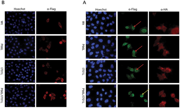
Figure 4. Effect of the NFATC1 missense SNPs on the cellular localization of the protein.
A- Immunofluorescence of HeLa cells transfected with plasmids encoding for the Wt NFATC1 and NFATC1 Mutants (P66L, I701L, P66L/I701L). The localization of NFATC1 was visualized using an anti-Flag antibody. Nuclei of cells were visualized using the Hoechst dye (blue color). Wt and NFATC1 mutants localized to the cytoplasm in the absence of PPP3CA (red color). (Magnification ×40). B- Immunofluorescence of HeLa cells transfected with plasmids encoding for the Wt NFATC1 and NFATC1 Mutants (P66L, I701L, P66L/I701L) co-transfected with PP3CA. The localization of NFATC1 was visualized using an anti-Flag antibody (red color) while PP3CA was visualized using anti-HA antibody (green color). Nuclei of cells were visualized using Hoechst dye (blue color). Most of the cells co-transfected with the double NFATC1 mutant were retained in the cytoplasm around the nuclear membrane, whereas in the other cases, the protein was totally translocated to the nucleus. (Magnification ×40). Yellow arrows indicate cytoplasmic (peri-nuclear) staining, while red arrows indicate nuclear staining.

