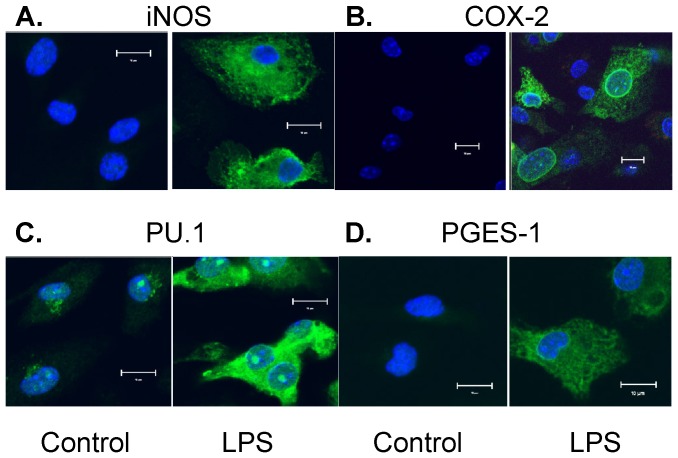
Figure 2. Confocol fluorescent microscopy detects the expression and intracellular distribution of iNOS/COX-2/PU.1/mPGES-1 induced by LPS treatment.
Primary cultured mouse BMDM was treated with or without 1 µg/ml LPS for 16 hrs. The protein expression of iNOS (A), COX-2 (B), PU.1 (C), and mPGES-1 (D) was determined by immunostaining followed by confocol fluorescent microscopy. In each sample group, BMDM was stained with the green fluorescent-labeled antibody for the targeted protein and the blue fluorescent-labeled DAPI for nucleus; the overlay image of each targeted protein and nucleus are shown (magnification: ×200, scale bar: 10 µM). LPS stimulation significantly increased cytosolic expression of iNOS, COX-2, mPGES-1 and PU.1 in BMDM, suggesting newly synthesized protein expression in cytosol. PU.1 also showed strong nuclear staining (i.e., nuclear translocation) after LPS treatment; whereas both COX-2 and mPGES-1 showed similar enhanced perinuclear localization in LPS-treated groups. The results shown are representative images from 3 independent experiments.

