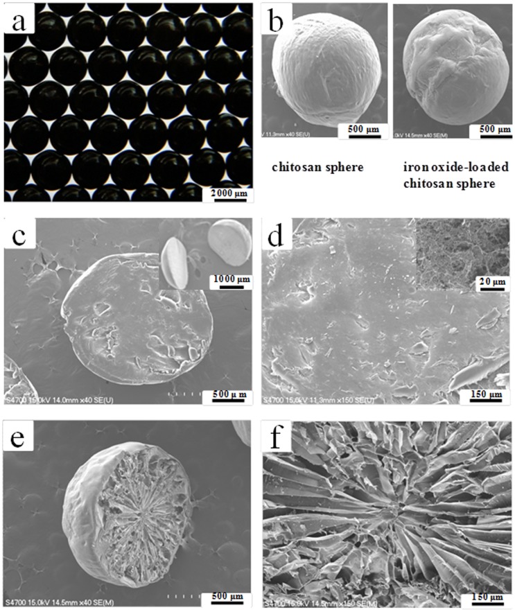
Figure 2. Morphologies of the synthesyzed chitosan spheres.
(a) An optical image of the iron oxide nanoparticles loaded chitosan spheres. (b) SEM images of chitosan spheres without (left) and with (right) iron oxide nanoparticles. (c) and (d) show the cross-section images of chitosan spheres (without iron oxide nanoparticles). Inset in (c) shows the sliced hemispheres of chitosan sphere. Inset in (d) shows details of the internal structure. (e) and (f) show the cross-section images of iron oxide nanoparticles loaded chitosan spheres.

