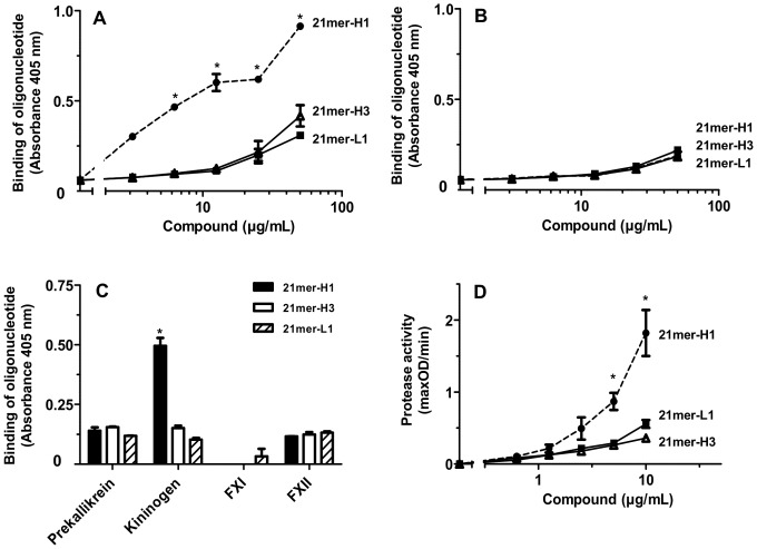
Figure 5. Binding of biotinylated DNA-oligonucleotides to different coagulation factors of the intrinsic coagulation pathway.
Microtiter plate wells were coated with 10 µg/mL each of (A) kininogen or (B) prekallikrein and binding of increasing concentrations of the biotinylated DNA-oligonucleotides 21mer-H1 (closed circles), 21mer-H3 (open triangles), 21mer-L1 (closed squares) was assessed. All data represent mean ± SD (n = 3; *p<0.05; 21mer-L1 and 21mer-H3 vs. 21mer-H1) of one representative experiment out of three independent ones. (C) Microtiter plate wells were coated with 10 µg/mL kininogen, factor XI (FXI) or factor XII (FXII) each and incubated with 25 µg/mL each of different biotinylated DNA-oligonucleotides: 21mer-H1 (black bars), 21mer-H3 (white bars) or 21mer-L (hatched bars). All data represent mean ± SD (n = 3) of one representative experiment out of three independent ones. (D) Increasing concentrations of the biotinylated DNA-oligonucleotides 21mer-H1 (closed circles), 21mer-H3 (open triangles) or 21mer-L1 (closed squares) were analyzed for prekallikrein auto-activation. All data represent mean ± SEM (n≥3; *p<0.05; 21mer-H1 vs. 21mer-H3).

