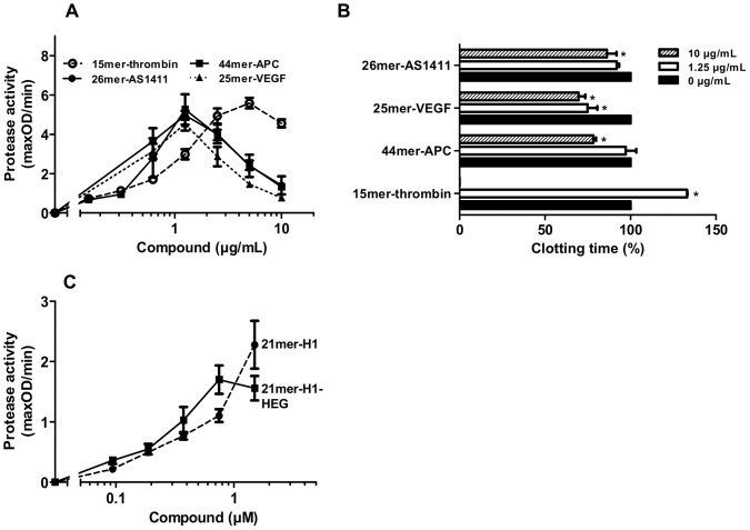
Figure 6. Influences of DNA-aptamers on the intrinsic coagulation pathway.
(A) The activation of prekallikrein was followed in the presence of increasing doses of the DNA-aptamers 15mer-thrombin (open circles, interrupted line), 44mer-APC (closed squares), 26mer-AS1411 (closed circles) or 25mer-VEGF (closed triangles, dotted line). (B) Turbidity clot-lysis assays were performed in the absence (black bars) or presence of 1.25 µg/mL (white bars) and 10 µg/mL (striped bars) of the DNA-aptamers 15mer-thrombin, 44mer-APC, 26mer-AS1411 or 25mer-VEGF, respectively. Coagulation was initiated by recalcification, clotting times were defined as respective time points of maximal absorbance. The clotting time of untreated plasma was defined as 100%. All data represent mean ± SEM (n = 3; *p<0.05; 1.25 µg/mL or 10 µg/mL vs. control). (C) The activation of prekallikrein was followed in the presence of increasing doses of the oligonucleotide 21mer-H1 (closed circles) and 21mer-H1-HEG (closed squares). All data represent mean ± SEM (n = 6).

