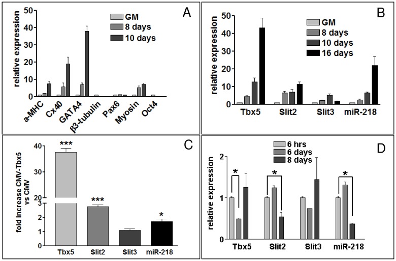
Figure 1. tbx5 and miR-218 are co-expressed in cardiomyocyte differentiation of P19CL6 cells.
A, qRT-PCR analysis of cardiac (α-MHC, Cx40, GATA4), muscle (Myosin), neural (Pax6, β-3-tubulin) and pluripotency (Oct4) markers in P19CL6 differentiating cells (8,10 days) compared to P19CL6 in growth medium (GM). B, qRT-PCR analysis of tbx5, slit2, slit3 and miR-218 relative expression in either expanding (GM) or differentiating (8,10,12 days) P19CL6 cells. (C-D), P19CL6 differentiating cells, 48 h after CMV-Tbx5 transfection compared to cells transfected with empty vector (C) or different times after Tbx5-siRNA transfection compared with cells transfected with siRNA-Ct (D). The timing course of the silencing experiment in (D) is described in the “Cell culture and transfection” section of methods. Results are standardized against GAPDH for genes, and against U6 for miRNAs.

