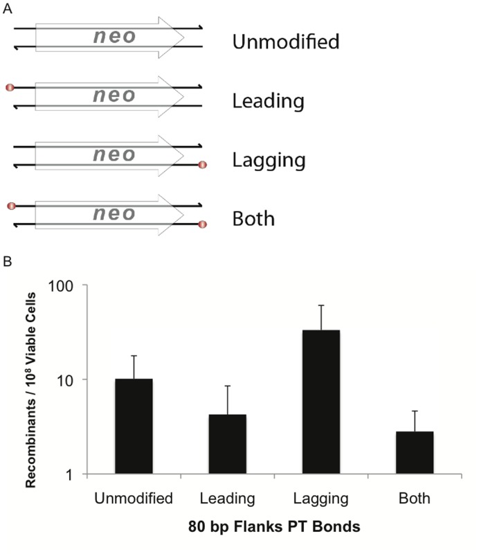
Figure 3. Phosphorothioate bonds on the matching lagging strand provide a modest increase in recombineering.
(A) Locations of the 5′ phosphorothioate bonds at the four terminal nucleotides are indicated as red spheres. Leading, Lagging and Both indicate the location of the 5′ phosphorothioate bonds on the respective strands of the substrates relative to the target location. (B) Recombination frequency for 80 nt flank protected substrates. 200 ng of each substrate was used in each electroporation. The results are the average of at least four independent replicates, error bars indicate standard deviation.

