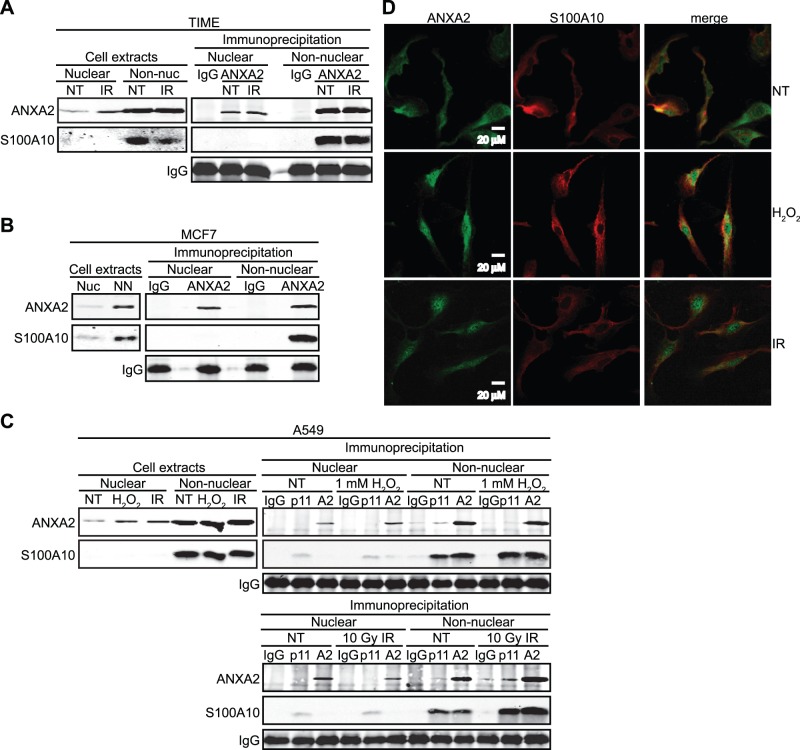
Figure 4. Nuclear annexin A2 is not associated with S100A10.
(A) TIME cells were either not treated (NT) or treated with 10 Gy IR for 2 hours; (B) MCF7 cells were treated with 10 Gy IR for 2 hours; (C) A549 cells were either not treated (NT) or treated with 1 mM H2O2 or 10 Gy IR for 2 hours as indicated. (A–C) Nuclear and non-nuclear fractions were prepared followed by immunoprecipitation with the antibodies indicated. Nuclear and non-nuclear fractions (cell extracts) and immunoprecipitates were subjected to SDS-PAGE and analyzed by western blotting with the antibodies indicated. (D) TIME cells were either not treated (NT) or treated with 0.5 mM H2O2 for 30 minutes or 10 Gy IR for 1 hour. Cells were subjected to immunocytochemistry analysis with the antibodies indicated and visualized by confocal microscopy. Scale bar is 20 µM.

