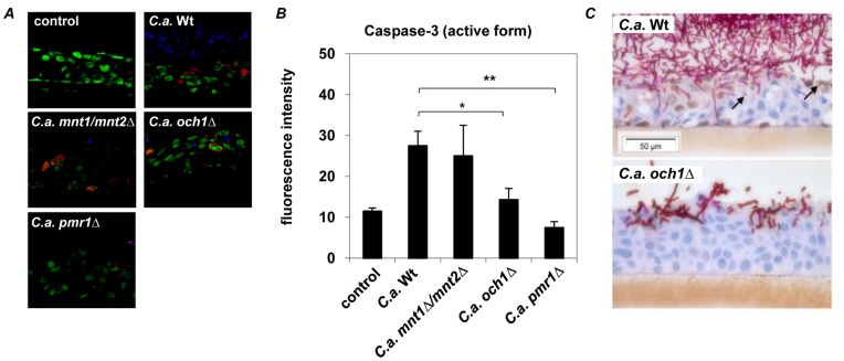
Figure 6. Cleaved (activated) caspase-3 expression in C. albicans infected oral RHE.
Oral RHE was infected with 2×10⁶ yeast cells from either C. albicans wild-type (SC5314), N-glycosylation (och1Δ), O-glycosylation (mnt1Δ/mnt2Δ) or N−/O-glycosylation (pmr1Δ) mutant strains for 4 h or 24 h. (A) Confocal microscopy of active caspase-3 expression (red) in RHEs after 4 h showed no caspase-3 induction in uninfected () or N−/O-glycosylation (pmr1Δ) mutant infected epithelium, whereas infection with C. albicans wild-type or O-glycosylation (mnt1Δ/mnt2Δ) mutant strain increased caspase-3 activation after 4 h of incubation. In N-glycosylation (och1Δ) mutant infected RHE reduced amounts of caspase-3 were observed. (B) Significantly reduced mean fluorescence intensity was observed in O-glycosylation (mnt1Δ/mnt2Δ) and N−/O-glycosylation (pmr1Δ) mutant infected RHEs compared to wild-type infected epithelium. (C) Cleaved caspase-3 expression (brown staining) is seen after 24 h in the nuclei of RHE cells adjacent to invading fungal hyphae and in apoptotic bodies (black arrows) in SC5314 but not Δoch1infected cultures. (A) Pictures shown are representative of three independent experiments; nuclei of epithelial cells are stained in green and C. albicans in blue. (B) Mean results of 10 analysed slides of three independent experiments, n = 3 (± SEM), *p<0.05, **p<0.01, 2-tailed paired Student’s t test.

