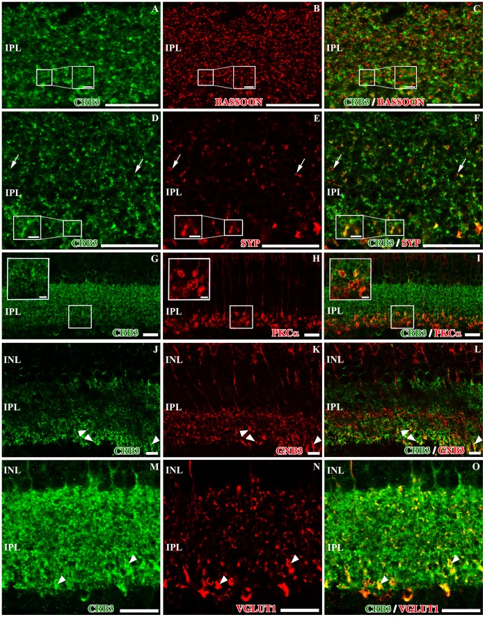
Figure 5. Localization of CRB3 in the inner plexiform layer of the adult mouse.
Double immunofluorescence for CRB3 (green) and bassoon (red in B–C), synaptophysin (SYP, red in E–F), PKCα (red in H–I), GNB3 (red in K–L) and VGLUT1 (red in N–O). A–F, CRB3 in the IPL surrounds the bassoon labeling (inset in A–C) and colocalizes with synaptophysin (inset in E–F), although some single labeled SYP positive profiles are found (arrows in D–F). G–L, in the IPL, PKCα is present in the axons of the rod bipolar cells (H–I), and GNB3 is present in the axons of a sub-population of cone bipolar cells (K–L). CRB3 colocalizes with both proteins in the innermost part of the IPL (insets in G–I and arrowheads in J–L). M–O, VGLUT1 is also present in the bipolar cells’ presynaptic terminals (N–O). All VGLUT1 positive processes colocalize with CRB3 (arrowheads in M–O), but not the opposite. Insets in A–I: Higher magnifications of the small squares. INL, inner nuclear layer; IPL, inner plexiform layer; GCL. Scale bars: 20 µm, 5 µm in inset (D–F) and 2 µm in inset (G–I).

