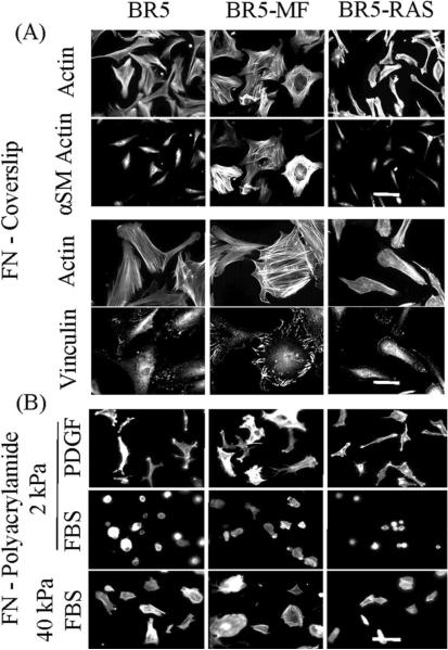
Figure 9.
Spreading of BR5, BR5 myofibroblasts, and oncogenic Ras-transformed BR5 cells on fibronectin-coated coverslips.
(A) BR5, BR5-MF and BR5-Ras fibroblasts as indicated were cultured 4 hr in PDGF-containing medium on fibronectin-coated coverslips. At the end of incubations, samples were fixed and stained for actin and α-smooth muscle actin and actin and vinculin. Scale bar, 100 μm (α-smooth muscle actin) and 50 μm (vinculin). (B) BR5, BR5-MF and BR5-Ras fibroblasts were cultured 4 hr in PDGF and FBS-containing medium as indicated on 2 and 40 kPa acrylamide gels with 50 μg/ml covalently crosslinked fibronectin. At the end of incubations, samples were fixed and stained for actin. Scale bar, 100 μm.

