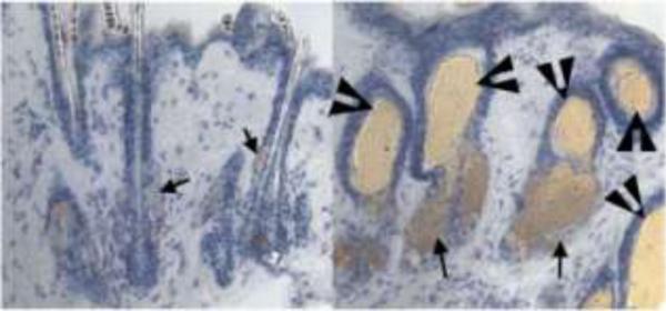
Fig. 3.

Skin phenotype of VDR-null mice. Skin from 8-month-old VDR-null mice (B) demonstrates lipid-laden dermal cysts (arrowheads) and an increase in sebaceous activity (arrows), relative to that seen in wildtype mice (A). Sections were stained for lipid with oil-red-O and counterstained with hematoxylin. Modified from [42]. Copyright 2007, National Academy of Sciences, U.S.A.
