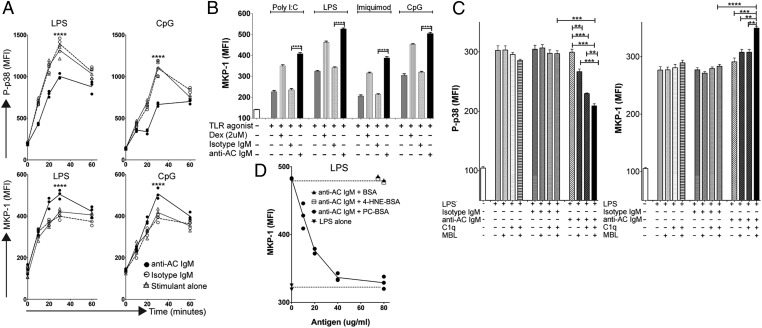
Fig. 2.
Kinetics and antigen specificity of anti-AC IgM inhibition of TLR-mediated p38 MAP kinase activation and MKP-1 induction. (A) Kinetic studies of intracellular levels of P-p38 (Upper panels) or MKP-1 (Lower panels) in DCs following exposure to anti-AC IgM and LPS (Left panels) or CpG (Right panels). Stimulations were performed in triplicates in serum-free media supplemented with C1q. Geometric mean fluorescence intensity (MFI) for MHCII-high DCs is shown (see Fig. S2 for gating). (B) MKP-1 induction by anti-AC IgM in response to agonists for diverse TLR. Results represent MFI mean ± SD for MHCII-high DCs from triplicate cultures after 30 min. (C) Inhibitory effects of anti-AC IgM on LPS-stimulated DCs requires C1q or MBL. Conditions are indicated for triplicate cultures at 30 min. (D) Concentration-dependent PC-antigen–specific inhibition of anti-AC IgM-mediated MKP-1 induction. DCs in serum-free media were cultured for 30 min without or with LPS under indicated conditions. Depicted values represent MFI from individual replicate cultures, with each condition performed in triplicate for MHCII-high DCs. C1q was used at 80 μg/mL and MBL at 20 μg/mL, based on pilot titration studies. Error bars indicate SD. **P < 0.01, ***P < 0.001, ****P < 0.0001.

