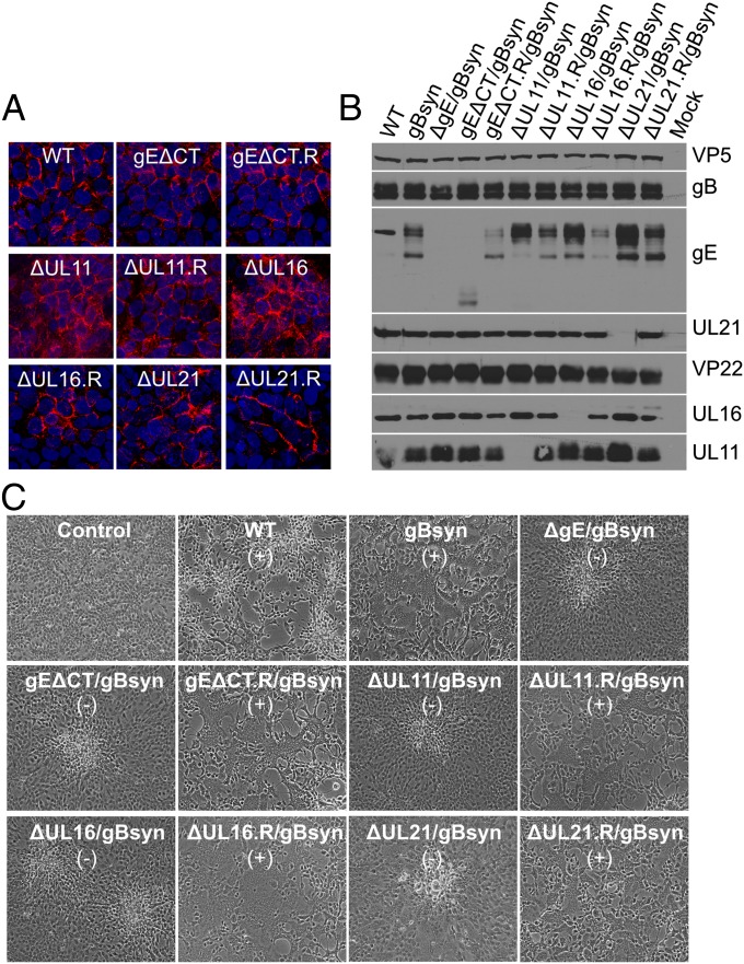
Fig. 6.
The Syn phenotype is lost in HaCaT cells even though gE accumulates on the cell surface when its binding partners are absent. (A) HaCaT cells grown on coverslips were infected with WT or the indicated mutant viruses at an MOI of 0.01. At 24 h after infection, the cells were fixed and reacted with a mouse monoclonal antibody (clone 3114) to gE before microscopy. (B) HaCaT cells were infected with WT or mutants viruses at an MOI of 5. At 18 to 24 h after infection, the cells were harvested, lysed in sample buffer, subjected to electrophoresis in denaturing gels, and reacted with the indicated antibodies. VP5 was used as a loading control. (C) HaCaT cells grown in six-well plates were infected with WT or Syn mutant viruses at low MOI. Virus-induced cytopathic effect was monitored daily and recorded by an inverted microscope. The presence (+) and absence (−) of syncytia is indicated.

