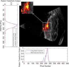
Fig. 1.
Coregistered photoacoustic and ultrasound image of a malignant ovary (the actual size of the coregistered image is ), displayed using different color maps, the figure also shows the Radon transform for 0 deg and 90 deg, both fitted with the Gaussian model to estimate the centroid of the area of interest. The photoacoustic image revealed clustered absorption distribution with higher intensity than that of benign cases.

