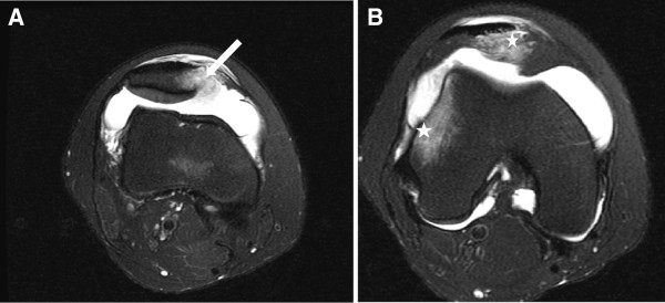
Figure 2.
An axial T2-weighted fast-spin-echo magnetic resonance imaging scan illustrates a eighteen year-old female sustaining a primary traumatic lateral dislocation of the patella while jumping. Complete avulsion of the medial patellofemoral ligament from its femoral insertion can be seen (arrow) (Figure 2A). The bone contusions (stars) (Figure 2B) of the lateral femoral condyle and the medial patellar facet are noted.

