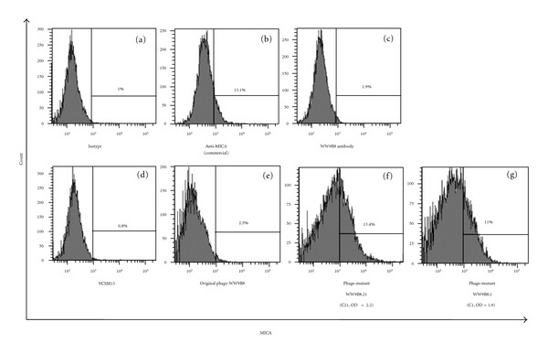
Figure 5.

Characterization of anti-MICA displayed phages against MICA-positive cells by flow cytometry. 293T cells (2 × 10⁵) were incubated with (a) mouse IgG1 isotype control PE (b) anti-human MICA antibody PE, (c) anti-MICA WW9B8 and followed by goat F(ab′)2 anti-mouse IgG-PE. (d, e, f, g) 293T cells (2 × 10⁵) were incubated with VCSM13 as negative control, original phages displaying Fab anti-MICA antibody (WW9B8), phages displaying mutant Fab WW9B8.21, phages displaying mutant Fab WW9B8.1 and followed by monoclonal antibody to M13 filamentous phage-FITC. The activities of phages displaying mutant WW9B8 are higher than the original phages carrying anti-MICA, WW9B8.
