Figure 2.
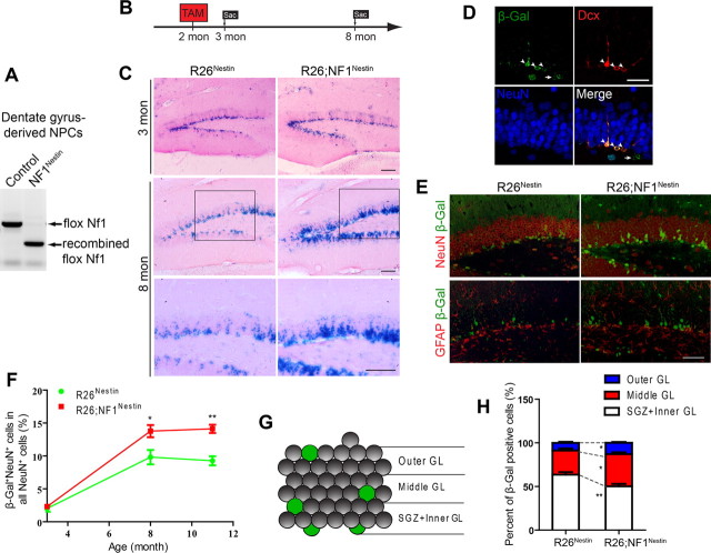
Ablation of Nf1 in adult NPCs in vivo. A, PCR demonstrating Nestin-CreERT2-mediated recombination of the flox Nf1 alleles in DG-derived adult NPCs. Mice were given tamoxifen at 2 months of age. NPCs were derived from DG at 3 months. B, Diagram of tamoxifen induction regimen. Mice were induced with tamoxifen at 2 months of age and killed at 3 months or 8 months of age. C, R26 reporter revealed enhanced long-term neurogenesis in the DG of NF1Nestin mice. Mice were given tamoxifen at 2 months and examined at 3 or 8 months of age. Images show representative X-Gal staining. Bottom show higher-magnification view of the boxed areas. TAM, tamoxifen. Scale bar, 100 μm. D, Coimmunostaining for β-Gal (green), Dcx (red), and NeuN (blue) at 3 months (1 month post-tamoxifen). Note that most recombined cells (β-Gal+) did not express NeuN, but colocalized with the immature neuronal marker (Dcx). Scale bar, 50 μm. E, In the DG of 8-month-old mice, adult-generated cells (β-Gal+) predominantly assumed neuronal fate (NeuN+). Images show representative double immunostaining for β-Gal (green) and NeuN (red, top) or GFAP (red, bottom). Scale bars, 100 μm. F, Quantification of β-Gal and NeuN double immunostaining revealed significant increase in the proportion of new-born neurons (β-Gal+NeuN+) in all NeuN+ granule neurons in NF1Nestin mice. ANOVA (GLM) revealed significant effects of time (F(2,25) = 101.6, p < 0.0001), genotype (F(1,25) = 21.49, p < 0.0001), and the interaction of the two (F(1,25) = 4.621, p < 0.0196). N = 4–6 for each genotype and age. G, H, Adult-born neurons (β-Gal+) in the DG of NF1Nestin mice exhibited increased migration into the middle and outer portions of the granular layers. GL, granular layer. N = 6 for each. Results are mean + SEM. *p < 0.05, **p < 0.01.

