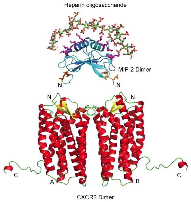
Figure 4.
GAG:MIP-2:CXCR2 interface model. The interface of heparin oligosaccharide, MIP-2 chemokine, and CXCR2 is shown in the model. The octadecameric heparin oligosaccharide encompasses the MIP-2 dimer over the helices. The monomers of MIP-2 are represented as ribbons (dark blue and light blue) with the side chains of heparin-interacting residues and the ELR motif as sticks (pink and orange, respectively). The heparin oligosaccharide MIP-2 model was aligned to MIP-2 of the MIP-2:CXCR2 complex (Figure 2) to obtain the tertiary model complex. The heparin oligosaccharide:MIP-2 interaction stabilizes MIP-2 dimer and does not interfere with binding to CXCR2.

