Figure 1.
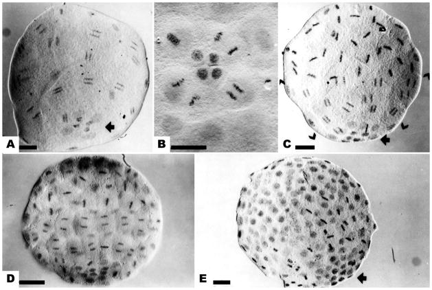
The mitotic gradient in cleavage stage embryos of Dendraster. (a) Mesomeres and macromeres synchronized in anaphase of the sixth division. Four small micromeres (SMi) and 4 large micromeres (LMi) at the vegetal pole have not entered mitosis (arrow). (b) The 6th division of the LMi. The SMi remain in interphase. (c) The 7th division of the mesomeres and macromeres. The mesomeres are in metaphase, the macromeres in anaphase (brackets), and the micromeres in the preceding interphase. Four SMi at the vegetal pole have not divided (arrow). (d) The eighth division of mesomeres and macromeres, while LMi complete their 7th division. (e) The ninth division of the macromeres, seen in metaphase. The micromeres can be identified as a group of small dense nuclei (arrow). Bar equals 25 μm.

