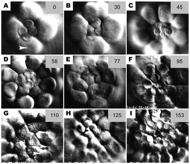
Figure 4.
Sequence of still photos from time-lapse video records of cleavage at the vegetal pole. (a) Late 16-cell stage. Micromere nuclei are intact whereas macromeres are beginning to furrow (arrowhead) (b) At the 5th cleavage, the micromeres have a slower cleavage schedule than the macromeres (c) The 32-cell stage, showing 4 small micromeres (SMi) atop 4 large micromeres (LMi); the 8 macromeres surround them. (d) The 4 LMi retain their nuclei while the macromeres undergo 6th cleavage. The SMi are above the plane or focus. (e) The 6th cleavage of the LMi. (f) The 60-cell stage showing 4 small and 8 LMi surrounded by the macromeres. (g) The 7th cleavage of the macromeres. Both small (arrowheads) and large (arrow) micromeres retain intact nuclei. Most micromere nuclei are not in focus in this photo, but all are intact. (h) The 6th cleavage of the SMi. LMi nuclei have broken down for the 7th cleavage. (i) The 8th cleavage of the macromeres. Note intact nuclei of the 24 micromeres. The numbers in the upper right of each frame are minutes elapsed from the first frames in the sequence at 17 °C.

