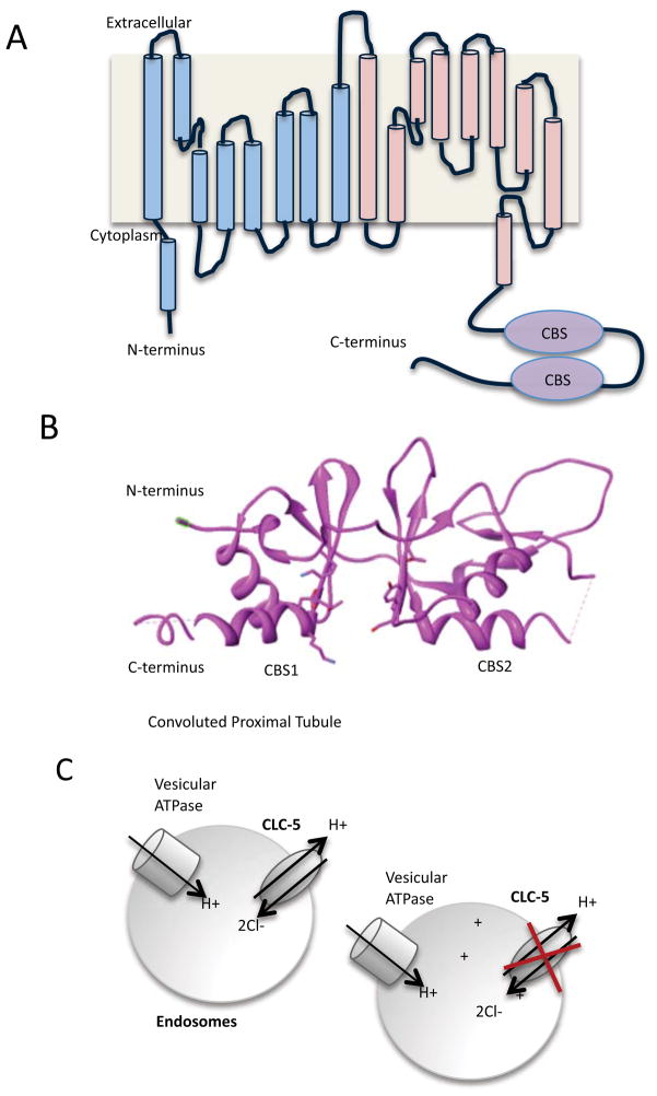
Figure 6.
ClC structure and function. A). Schematic arrangement of the α helices that are in mammalian ClC. It is believed that there are 10–12 transmembranes in ClC, however, not all helices span the membrane. The first 9 α helices (shaded blue) and the second 9 α helices (shaded pink) are similar in structure, though there is little homology at the amino acid level. Note the presence of the two cystathionine β-synthetase binding sites domains in the C-terminal tail. B). Structure of the cystathionine β-synthetase binding sites by X-ray crystallography30. A cleft between the two CBS sites binds the nucleotides. PDB accession code: 2J9L. Structure was modified the UCSF Chimera package142. C). ClC-5 acts to transport H+ and Cl− in endosomes, to counteract the activity of vesicular ATPase in the convoluted proximal tubule. Loss of ClC-5 function leads to the acidification of the endosome.

