
Figure 3.
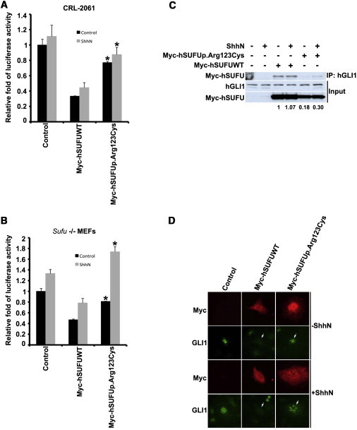
The Effect of the p.Arg123Cys Altered SUFU on Hedgehog Signaling Activity
(A and B) The p.Arg123Cys altered SUFU has decreased activity. Human rhabdomyosarcoma cells (CRL-2061) and Sufu-deficient (Sufu−/−) MEFs were transfected with Myc-tagged wild-type (Myc-SUFUWT) or altered (Myc-SUFUp.Arg123Cys) SUFU constructs, an Hh-pathway-specific reporter, and a control reporter. The cells were incubated with and without ShhN-conditioned medium, and luciferase activity was measured. Note that Myc-SUFU suppresses Hh-pathway activity in both human and mouse cell lines. ∗p < 0.05 compared with Myc-hSUFUWT (statistical analysis was done with a t test). Error bars indicate one standard deviation (duplicate samples).
(C) The p.Arg123Cys altered SUFU binds less GLI1 than does the wild-type SUFU. CRL-2061 cell lysates used in luciferase reporter assay were subjected to pull-down immunoblot analysis (IP: GLI1; WB: Myc and GLI1). In the top panel, Myc-SUFU is pulled down with antibodies against GLI1. Input of GLI1 and Myc-tagged SUFU constructs is presented in the middle and bottom panels, respectively. Quantification of GLI1 binding relative to the Myc-SUFU protein level is shown below the panel (quantitative protein analysis was performed with the Single Dimensional Electrophoretic Gel Analysis program from the ImageJ software package v.1.46g).
(D) Subcellular localization of p.Arg123Cys altered SUFU in Sufu−/− MEFs. Note that the Gli1 nuclear localization was not affected by the human SUFU mutant (Myc-SUFUp.Arg123Cys) compared with the wild-type SUFU (Myc-SUFUWT), as indicated by arrows.