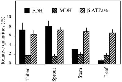
Figure 2.
Relative contents of FDH, MDH, and β-ATPase proteins in potato mitochondria isolated from different tissues, as estimated by quantitation of the corresponding spots on Coomassie blue-stained two-dimensional gels, as described in Methods. The relative abundance of each spot was expressed as a percentage of the total number of protein spots detected on the gels. The figures are an average of five quantitations performed on five gels resulting from five distinct mitochondrial preparations.

