
Figure 3.
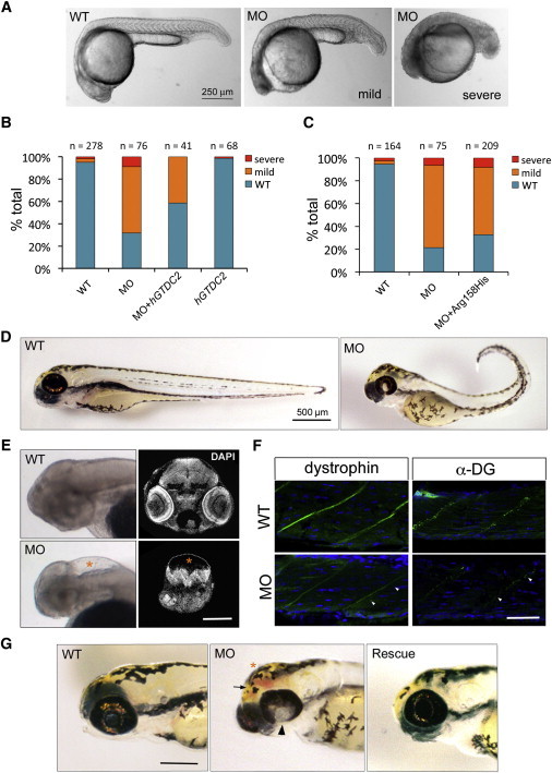
Loss of gtdc2 in the Zebrafish Embryo Recapitulates Phenotypes Observed in WWS
(A) Mild (center) and severe (right) phenotypes observed in the 1 dpf zebrafish embryo after injection of a MO designed against the start site of gtdc2.
(B) Phenotypes observed at 1 dpf can be rescued by coinjection of the gtdc2 MO and 150 pg of GTDC2 mRNA, whereas no effect is observed by injection of the mRNA alone (hGTDC2).
(C) Coinjection of 150 pg of GTDC2 mRNA containing the p.Arg158His alteration is not able to rescue the morphant phenotype. Representative experiments are shown.
(D) Surviving morphants at 3 dpf show several developmental defects: reduced length, bent tail, failure of ventral retinal fusion, and dysmorphic head shape.
(E) 3 dpf morphants also display hydrocephalus (lower left panel: whole-mount of nonpigmented larva [asterisk]) and ventral compression of the remaining brain tissue (lower right panel: DAPI staining on histological section at a location matching the wild-type [WT] section above). The scale bar represents 200 μm.
(F) Immunohistochemistry in sections of the WT and morphant muscle shows that expression of both dystrophin (green, left) and glycosylated dystroglycan (green, right) are reduced at the myosepta (marked by arrowheads). Nuclei are labeled in DAPI (blue). The scale bar represents 50 μm.
(G) Rescue with GTDC2 mRNA (on right) ameliorates the most severe head phenotypes observed in the morphant (center). These phenotypes are failure of retinal fusion (arrowhead), hydrocephalus (asterisk), and hemorrhage (arrow). The scale bar represents 200 μm.