Fig. 2.
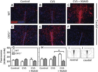
Antiinflammatory treatment prevents chronic stress-induced increases in GFAP in the PVN of stress-sensitive CRF2−/− mice. A–F, Representative immunofluorescence images (×10 magnification) of the astrocyte-specific cytoskeletal protein, GFAP (red), counterstained with DAPI (nuclei, blue) in the caudal PVN of (A–C) WT and (D–F) CRF2−/− mice for control (A and D), after exposure to CVS (B and E), or CVS + NSAID (C and F). Bar graphs illustrate semiquantitative analysis of GFAP immunofluorescence. G, No differences between groups were detected with CVS on GFAP in the rostral PVN. H, In the caudal PVN, NSAID treatment increased GFAP in WT but decreased GFAP in CRF2−/− mice. Atlas images illustrating the brain sections analyzed for rostral (I) and caudal (J) PVN, adapted from the mouse atlas (26). Data are presented as parameter estimates of the best-fit model + observed sd of respective groups (n = 3–4). *, P < 0.05.

