Fig. 3.
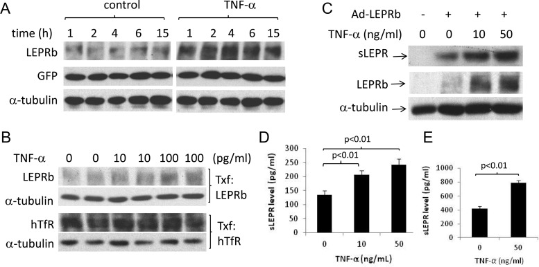
TNF-α stimulates LEPRb protein expression and sLEPR release in multiple cell types. A, Western blot analysis of LEPRb, GFP, and α-tubulin levels in cell lysates. N2a cells were cotransfected with pcDNA plasmids carrying LEPRb and GFP cDNA. Transfected cells were then treated with vehicle or 1 ng/ml TNF-α for 1–15 h before the analysis. B, Western blot analysis of LEPRb, hTfR, and α-tubulin levels in cell lysates. N2a cells were transfected with the LEPRb expression plasmid or with the hTfR expression plasmid. Transfected cells were then treated with TNF at 0, 10, or 100 pg/ml for 1 h. LEPRb-transfected cells were analyzed for LEPRb and α-tubulin, and hTfR-transfected cells for hTfR and α-tubulin as indicated. C, Western blot analysis of LEPRb and α-tubulin in cell lysates and sLEPR in CM. LEPRb-transfected N2a cells were treated for 6 h with 0, 10, and 50 ng/ml TNF-α before the analysis. Lysates and CM from untransfected N2a cells were analyzed in parallel as controls. D, ELISA analysis of sLEPR levels in the CM of GT1-7 hypothalamic neuronal cells. Ad-LEPRb-infected GT1-7 cells were incubated for 16 h with 0, 10, and 50 ng/ml TNF-α before CM were collected for ELISA analysis. Endogenously produced mouse sLEPR, if any, was not detected by ELISA analysis in the CM of uninfected GT1-7 cells. E, ELISA analysis of sLEPR levels in the CM of HepG2 cells. LEPRb-transfected HepG2 cells were treated for 15 h with vehicle or with 50 ng/ml TNF-α before CM were analyzed by ELISA. No sLEPR in the CM of untransfected HepG2 cells was detected by ELISA.

