Fig. 5.
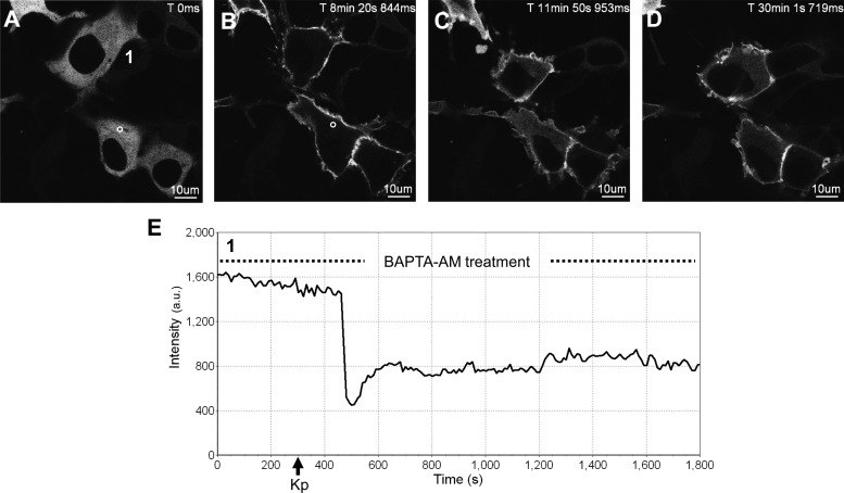
The effect of BAPTA-AM pretreatment on the spatiotemporal characteristics of GFP-PKC-βII in HEK 293 cells in response to KISS1R activation. A–D, Representative images selected from a time series of laser-scanning confocal microscopic images showing the plasma membrane and cytosolic localization of GFP-PKC-βII in KISS1R-expressing HEK 293 cells pretreated with 50 μm BAPTA-AM (see dashed line in panel E) followed by 100 nm Kp-10 approximately 250 sec later (see solid black arrow on x-axis of panel E) in the continued presence of BAPTA-AM (see dashed line in panel E). Cytosolic region of interest in a cell (shown by small white circle in panel A) was analyzed quantitatively by determining the changes in GFP fluorescence over time. These changes are presented graphically (panel E). Note: although cells were imaged for approximately 1 h, due to their movement over this period, a given region of interest could only be analyzed for subperiods of time. As a result, we present a region of interest that represents the first 30 min of imaging (panel G). These studies were conducted five independent times, and representative images from one independent experiment are shown. ms, Millisecond; s, second.

