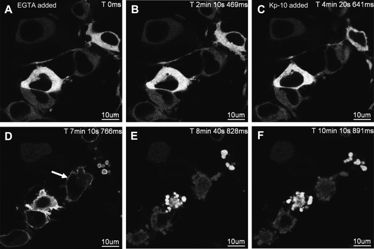
Fig. 6.
The effect of EGTA pretreatment on the spatiotemporal characteristics of GFP-PKC-βII in HEK 293 cells in response to KISS1R activation. A–F, Representative images selected from a time series of laser-scanning confocal microscopic images showing the plasma membrane and cytosolic localization of GFP-PKC-βII in KISS1R-expressing HEK 293 cells pretreated with 2.5 mm EGTA followed by 100 nm Kp-10 approximately 4 min later in the continued presence of EGTA. White solid arrow shows transient translocation of GFP-PKC-βII (panel D). Prolonged exposure to EGTA caused cells to lift due to the chelation of extracellular Ca2+; as a result, cells could only be imaged for up to 10–25 min after EGTA treatment. These studies were conducted five independent times, and representative images from one independent experiment are shown. ms, Millisecond; s, second.

