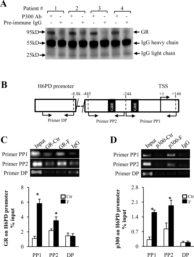
Fig. 6.
A, Coimmunoprecipitation assay showing the detection of GR in the nuclear protein complex precipitated by p300 antibody upon stimulation of human amnion fibroblasts with cortisol (F; 1 μm). Preimmune IgG served as negative control. B, Diagram illustrating the positions of putative GREs and primer sets used for the ChIP assay. C and D, Top panels are the representative gel images showing the abundance of PCR products, and bottom panels are the mean data of qRT-PCR showing the enrichment of p300, GR to H6PD promoter upon cortisol (F; 1 μm) stimulation of human amnion fibroblasts. Preimmune IgG served as negative control (n = 3–4). *, P < 0.05 vs. control (CTR). TSS, Transcription start site; PP1, proximal primer set 1; PP2, proximal primer set 2; DP, distal primer set.

