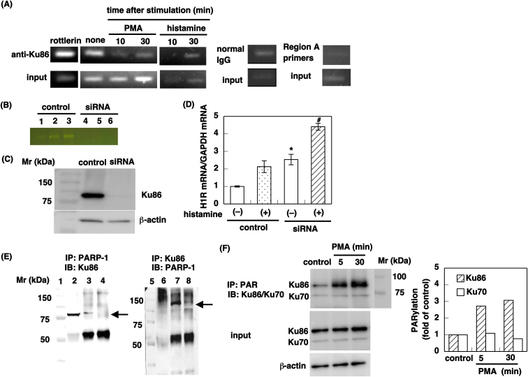
Figure 5. Dissociation of Ku86 from the promoter in response to stimulation activates the H1R promoter activity.
(A) Chip assay. HeLa cells were serum-starved for 24 h and stimulated with or without 100 nM of PMA or 100 μM of histamine for the times indicated. DNA–protein complexes were immunoprecipitated with anti-Ku86 and PCR was performed to amplify the region B1 promoter. For the control, HeLa cells were pretreated with 10 μM of rottlerin 1 h before PMA stimulation to completely eliminate H1R signaling20. For input, 1% of the extract was used. (B-D) Knockdown study. HeLa cells were reverse-transfected with 10 μM Ku86-specific siRNA (represented as siRNA) or 10 μM control siRNA (control). Forty-eight hours after transfection, the cells were starved for 24 h followed by treatment with histamine (100 μM). Expression of Ku86 mRNA (B) and protein (C) was analyzed. In B, total RNA was prepared from 3 individual dishes. Lanes 1, 2, and 3: control; lanes 4, 5, and 6: Ku86-specific siRNA. In D, The H1R mRNA levels were determined by real-time quantitative RT-PCR. Data are presented as the means ± S.E.M. (n = 3). *, p < 0.05 versus control, histamine (–); #, p < 0.05 versus control, histamine (+). (E) PMA stimulation induces dissociation of Ku86 from PARP-1. Immunoprecipitation assay was conducted as described in Methods. Lanes 1 and 5, marker; lanes 2 and 6, input (1% of the extract); Lanes 3 and 4, the extract incubated with anti-PARP-1 antibody and immunoprecipitated proteins were SDS-PAGE separated and analyzed with anti-Ku86 antibody. Lanes 7 and 8, the extract incubated with anti-Ku86 antibody and immunoprecipitated proteins analyzed with anti-PARP-1 antibody. Lanes 3 and 7, control; Lanes 4 and 8, PMA stimulation. For the control, HeLa cells were pretreated with 10 μM of rottlerin 1 h before PMA stimulation. Arrows indicate Ku86 (left panel) and PARP-1 (right panel). (F) PMA stimulation induces PARylation of Ku86 by PARP-1. Left panel: HeLa cells were serum-starved for 24 h and stimulated with or without 100 nM of PMA for the time indicated. Immunoprecipitated proteins by anti-poly(ADP-ribose) antibody were analyzed with anti-Ku86/Ku70 antibodies. Right panel: Quantification of PARylation of Ku86 and Ku70. Densitometric analysis was performed using ImageJ software.

