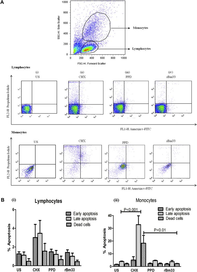
Fig. 5.
Flow cytometric analysis of apoptosis (A) Representative distributions of the fluorescence intensity of Annexin V-FITC and propidium iodide binding of human lymphocytes and monocytes that were flow cytometrically gated using the forward- and side-light scatters in human Peripheral Blood Mononuclear Cells (PBMCs). PBMCs cultured in the presence of medium only, (i) Unstimulated cells (US),(ii) Cycloheximide (CHX, 100 μM), (iii) Purified Protein Derivative from M. tuberculosis (PPD,10 μg/ml), (iv) Recombinant Bm33 (rBm33,10 μg/ml). (B) Quantitative analysis of apoptosis by FACS: (i) Percentage of apoptosis in lymphocyte population of PBMCs, (ii) Percentage of apoptosis in monocyte population of PBMCs. Values are mean ± S.D obtained from five independent experiments.

