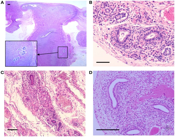
Figure 2.
Hematoxylin and Eosin stained sections with areas of ectopic endometrial glands and embryonic ducts. Histological appearance of ectopic glands and stroma observed (insert at higher magnification) in fetal uterine wall (A). Presence of embryonic ducts located in the broad ligament (B), under the fallopian tube serosa (C), and ducts located in the ovarian ligament (D). Note presence of a stromal component surrounding the duct residues in (A–D). Scale bars, 100 μm.

