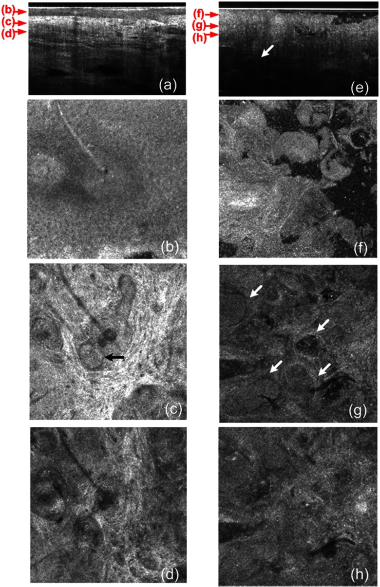
Fig. 4.
(a) A cross-sectional GD-OCM image of a normal nose skin; (b), (c), and (d) are the en face images at the corresponding depth indicated with the red arrows in Fig. 4(a), respectively; (e) a cross-sectional GD-OCM image of BCC located in nose skin; (f), (g), and (h) are the en face images at the corresponding depth indicated with the red arrows in Fig. 4(e), respectively. The size of the cross-sectional images (a) and (e) is , and the size of the en face images is .

