
Fig. 3.
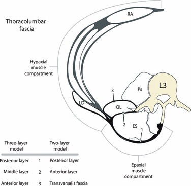
This is a tracing of the hypaxial and epaxial myofascial compartments, illustrating the comparison between the two-layered and three-layered models of the TLF. The latissimus dorsi (LD) is seen lying on the external wall of the hypaxial compartment and extending over the epaxial compartment to reach its attachments on the midline. In doing so, the aponeurosis of the LD contributes to the superficial lamina of the PLF.