
Fig. 7.
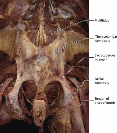
This is a posterior view of the lumbosacral region following removal of the gluteus maximus and the erector spinae muscles. Multifidus lumborum is seen inserting into the TLC. The composite extends caudally to cover the sacrotuberous ligaments and reach the ischial tuberosities.