
Fig. 14.
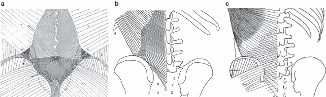
A comparison between drawings of three studies of the superficial lamina of the PLF: (a) Vleeming et al.; (b) Bogduk et al.; (c) Barker et al. (a–c) The same fiber direction of the superficial lamina. (a and c) A crosshatched appearance and the connections to the gluteus maximus fascia. (a) Along with variation in fiber direction there are changes in fiber density in the superficial lamina as well. Where the abdominal muscles join the paraspinal muscles, the orientation of the fibers change and they become denser. From the level of L4 to the lower part of the sacrum, the fiber density markedly increases. This density change corresponds to the area where the different fascial layers fuse to form the TLC.