
Fig. 15.
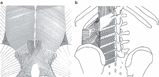
A comparison between drawings of two studies of the deep lamina of the PLF: (a) Vleeming et al.; (b) Bogduk et al. (a and b) The same fiber direction; however, in (b) the dense parts of the deep lamina are coined as accessory ligaments. (b) The lateral raphe is indicated as a dotted vertical line, indicating the area where the abdominal muscles join the paraspinal muscles. (a) An increase of density in the same area. More caudally, it can be noticed that the sacrotuberous ligament partially fuses to the deep lamina. The fiber characteristics show increased density and an altered pattern in the region over the sacrum. This pattern is another indication that the various layers of the TLF and aponeurosis fuse into the TLC, as referred to in the text.