
Fig. 9.
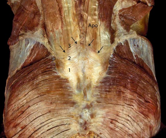
Components of the composite over the lumbosacral spine. The superficial lamina of the PLF (1), which is dissected at (2) where the deep lamina of the PLF becomes visible. The deep lamina (2) is dissected and the aponeurosis tendon of the erector trunci and multifidi becomes visible (APO). (With permission from the Willard Carreiro collection.)