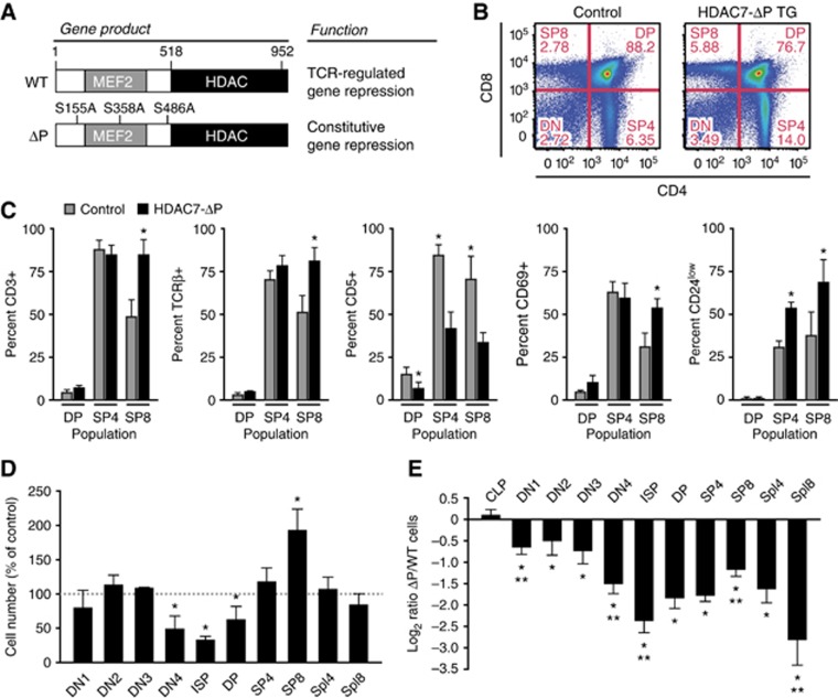
Figure 1.
Expression of HDAC7-ΔP alters T-cell development. (A) Locations of S-A mutations in HDAC7-ΔP and the resultant alteration of HDAC7 function. (B) Representative scatter plots showing change in thymic CD4 and CD8 expression profiles in WT control (left) and HDAC7-ΔP TG (right) mice. (C) Effect of HDAC7-ΔP on expression of CD3ε, TCRβ, CD5, CD69, and CD24 in DP, CD4 SP, and CD8 SP thymocytes, gated as shown in Supplementary Figure S1C. Bars show percent ±s.d. for five control/HDAC7-ΔP TG littermate pairs. *P=6 × 10⁻⁵−0.014, two-tailed paired T-test. (D) Percent relative to WT littermate controls of cells present at the indicated developmental stages in HDAC7-ΔP TG mice. Data are from six control/HDAC7-ΔP TG littermate pairs±s.e.m. *P=0.0057−0.039, two-tailed paired T-test. (E) Log2 ratio of WT/ HDAC7-ΔP cells present at indicated stages in radiation chimeras reconstituted with equal amounts of wild-type control and HDAC7-ΔP bone marrow. Data are from eight animals engrafted with WT and transgenic bone marrow at a 1:1 ratio, ±s.e.m. *P=2.7 × 10⁻⁹−0.026 versus WT cells, two-tailed T-test; **P=0.001−0.04 versus previous stage, two-tailed paired t-test. (D, E) CLP, common lymphoid progenitor; DN1-DN4, double-negative 1–4 thymocytes; ISP, immature single-positive; DP, CD4/CD8 double-positive; SP4, 8, CD4, 8 single-positive; Spl4, 8, spleen CD4, CD8 T cells.

