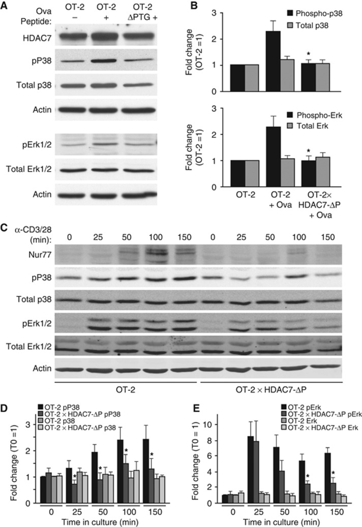
Figure 4.
HDAC7-ΔP blocks MAP kinase activation after strong TCR engagement. (A) Representative western blot showing phospho-p38, total p38, phospho-Erk1/2, total Erk1/2, HDAC7, and Actin expression for unstimulated OT-2 DP thymocytes (left), OT-2 DP thymocytes 3 h after agonist peptide injection (middle), and HDAC7-ΔP X OT-2 DP thymocytes 3 h after agonist peptide injection (right). (B) Quantification by optical densitometry of phospho- and total p38 (top), and Erk (bottom) for DP thymocytes from four sets of animals prepared as in (A). *P=0.008–0.028 for stimulated OT-2 X HDAC7-ΔP versus stimulated littermate OT-2 thymocytes, two-tailed T-test. (C) Representative western blots showing expression of Nur77, phospho-P38, total P38, phospho-Erk, total Erk, and actin for OT-2 and OT-2 X HDAC7-ΔP DP thymocytes stimulated ex vivo with α-CD3+α-CD28 for the indicated times. (D) Fluorimetric quantification of phospho- and total P38 in OT-2 and OT-2 X HDAC7-ΔP DP thymocytes stimulated with α-CD3+α-CD28 for the indicated times. *P=0.0006–0.027, paired two-tailed T-test. (E) Fluorimetric quantification of phospho- and total Erk in OT-2 and OT-2 X HDAC7-ΔP DP thymocytes stimulated with αCD3+αCD28 for the indicated times. *P=0.0012–0.013, paired two-tailed T-test. Figure source data can be found with the Supplementary data.

