Figure 5.
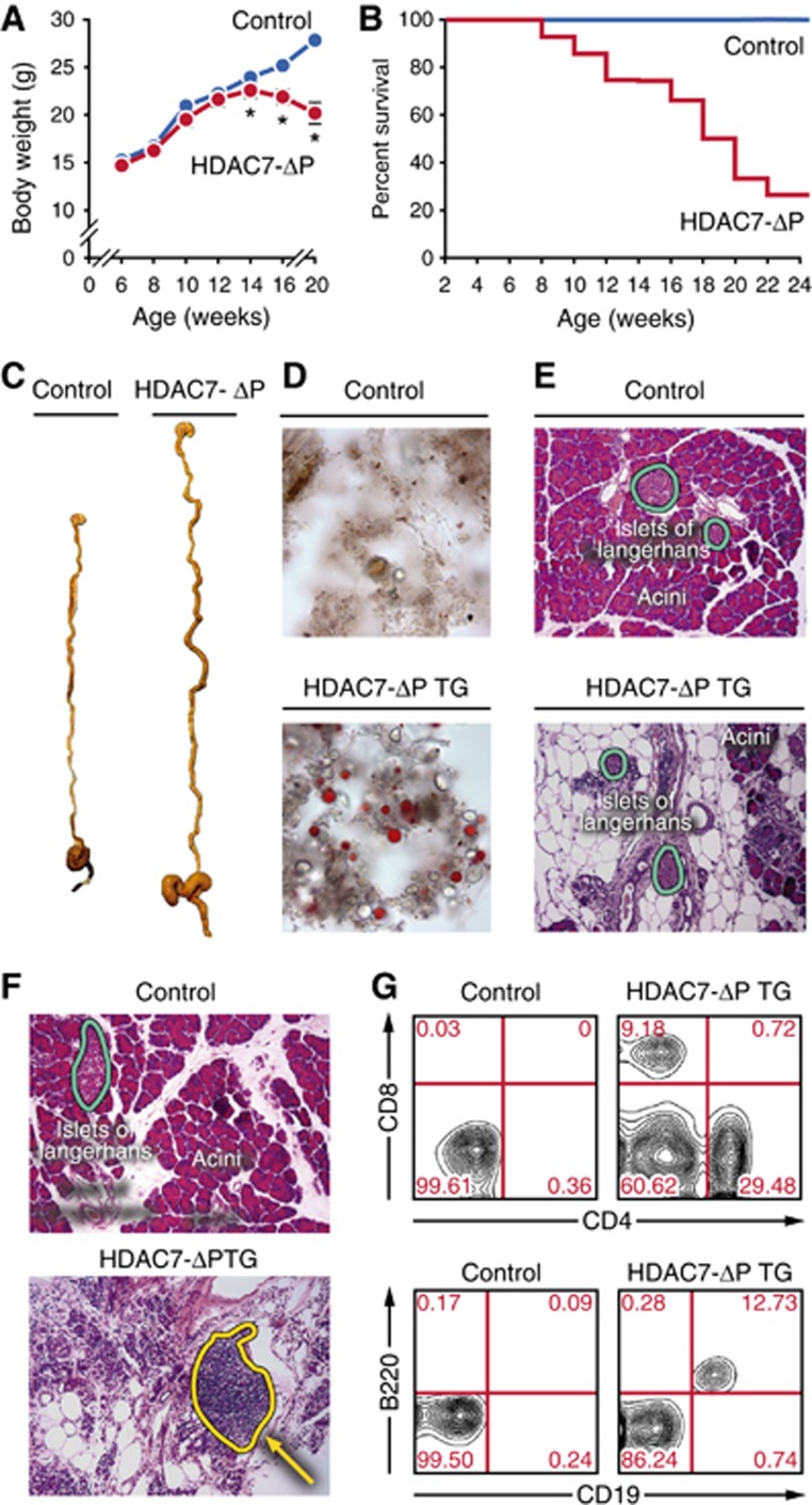
Thymic expression of HDAC7-ΔP causes lethal exocrine pancreatitis. (A) Body weight in grams of WT and HDAC7-ΔP TG animals between 6 and 20 weeks of age. Values are mean±s.e.m. of eight transgenic animals and corresponding WT littermates. *P<0.01, two-tailed T-test. (B) Percent survival of cohorts of eight HDAC7-ΔP TG and eight WT littermate animals from 2 to 24 weeks of age. (C) Representative gross specimens of WT littermate (left) and HDAC7-ΔP TG (right) digestive systems from animals 14 weeks of age. (D) Representative Sudan IV-stained faecal smear micrographs from WT littermate control (top) and HDAC7-ΔP TG (bottom) mice, demonstrating steatorrhea in HDAC7-ΔP TG animals. (E) Representative haematoxylin and eosin stained sections of WT (top) and HDAC7-ΔP TG (bottom) pancreas, showing massive destruction of acinar tissue but not islets in HDAC7-ΔP TG animals. (F) Representative haematoxylin and eosin stained sections of wild-type littermate control (top) and HDAC7-ΔP TG (bottom) pancreas, showing extensive immune infiltrate in HDAC7-ΔP TG specimen (dotted yellow outline). (G) Representative scatter plots measuring CD4 and CD8 (top) or CD19 and B220 expression (bottom) in pancreatic infiltrate cells from wild-type littermate control (left) and HDAC7-ΔP TG (right) mice.

